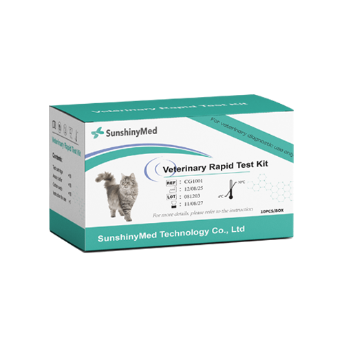


Feline Panleukopenia Virus Antigen Rapid Test
The SunGold™ Feline Panleukopenia Virus Antigen Rapid Test uses colloidal gold immunochromatography technology to identify the feline panleukopenia virus (feline panleukopenia virus) antigen through specific monoclonal antibodies, and forms a visual detection line on the test card after binding with the target antigen in the sample, which is used to quickly determine whether the cat is infected with the virus.
Production Features
Simple sampling
Easy to operate
Results in 10 minutes
Reduce testing costs
Product Parameters
| Production Name | SunGoldTM Feline Panleukopenia Virus Antigen Rapid Test |
| Usage | Veterinary Diagnostics |
| Detection Method | Chromatographic Immunoassay/Lateral Flow Immunoassay |
| Target Analyte | Feline Panleukopenia Virus Antigen |
| Specimen Type | Feces |
| Storage Temperature | 4-30°C |
| Result Time | 5-10 Minutes |
| Packaging Specification | Individually sealed, 10 tests in total. |
| Format | Cassette |
| Shelf Life | Up to Expiration Date Indicated on Package |
Product Performance
| Sensitivity | 98.00% |
| Specificity | 97.50% |
| Accuracy | 97.71% |
Description
SunGold™ Feline Panleukopenia Virus Antigen Rapid Test is mainly used in animal hospitals, pet clinics, stray cat rescue stations and other places, and is suitable for early screening of cats with symptoms such as fever, vomiting, diarrhea, and a sudden decrease in white blood cells. The background of the project development is that feline panleukopenia is a highly contact-transmitted viral disease, especially in kittens. The mortality rate is high. Early identification is crucial to prevent and control transmission and improve the success rate of treatment. The SunGold™ Feline Panleukopenia Virus Antigen Rapid Test uses a chromatographic immunoassay method and provides results within 5–10 minutes after sample application, making it fast and convenient.
How to use?
Check the product contents and make sure the test operation is under the room temperature (15–30℃) before testing.
Unseal the extraction tube containing the buffer.Place the extraction tube in the workstation.
Squeeze the upper air bag to absorb the sample (whole blood/serum/plasma) . Please make sure some sample getting into the lower air bag and there is no bubbles in the lower tube. Then press the upper air bag to transfer the sample (about 75 μl) left in the lower tube into the buffer.
Cover the tube, Jiggle the extraction tube until the specimen and the buffer are mixed completely.
Take the test device out of the aluminum foil bag, and place it on a clean, flat table. Add three drops (about 90 μL) of the mixed sample vertically into the specimen well (S) of the test device.
Interpretation of Results
Positive (+): The presence of both C line and T line, regardless of T line being strong or faint.
Negative (-): Only clear C line appears.
Invalid: No colored line appears in C region, regardless of T line's appearance.
Materials provided:
10 test cards
10 vials of assay buffer
10 bags of swab stick
1 package insert


