
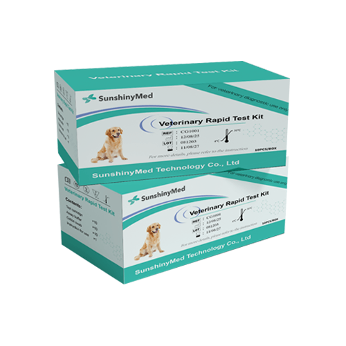

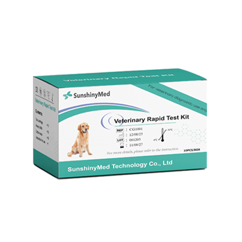

Babesia Gibsoni Antibody Rapid Test
The SunGoldTM Babesia Gibsoni Antibody Rapid Test employs colloidal gold immunochromatography (GICA) technology to rapidly detect antibodies to Giardia parasites in serum, plasma, or whole blood through specific capture. The experimental principle is based on the double-antigen sandwich method. Antibodies in the sample bind with colloidal gold-labeled antigens to form complexes, which are then captured by fixed antigens at the test line and undergo color development.
Production Features
Simple sampling
Easy to operate
Results in 10 minutes
Reduce testing costs
Product Parameters
| Production Name | SunGoldTM Babesia Gibsoni Antibody Rapid Test |
| Usage | Veterinary Diagnostics |
| Detection Method | Chromatographic Immunoassay/Lateral Flow Immunoassay |
| Target Analyte | Babesia Gibsoni Antibody |
| Specimen Type | whole blood, serum or plasma sample |
| Storage Temperature | 4-30°C |
| Result Time | 5-10 Minutes |
| Packaging Specification | Individually sealed, 10 tests in total. |
| Format | Cassette |
| Shelf Life | Up to Expiration Date Indicated on Package |
Product Performance
| Sensitivity | 97.33% |
| Specificity | 98% |
| Accuracy | 97.67% |
Description
Qualitative results of Babesia Gibsoni can be obtained within 10 minutes, with simple operation requiring no specialized equipment. This product is suitable for clinical diagnosis in veterinary clinics, health screening in kennels, and animal disease control monitoring, effectively distinguishing between acute and chronic infections and assessing treatment efficacy. Babesia gibbsii is an important blood parasite in dogs, capable of causing severe hemolytic anemia and even death. Traditional microscopy has low sensitivity, while PCR testing is costly. This test kit provides clinicians with a highly specific (>97%) and highly sensitive (>95%) on-site rapid screening solution, significantly improving diagnostic efficiency and aiding in the prevention and control of tick-borne diseases.
How to use?
Check the product contents and make sure the test operation is under the room temperature (15–30℃) before testing.
Unseal the extraction tube containing the buffer.Place the extraction tube in the workstation.
Squeeze the upper air bag to absorb the sample (whole blood/serum/plasma) . Please make sure some sample getting into the lower air bag and there is no bubbles in the lower tube. Then press the upper air bag to transfer the sample (about 75 μl) left in the lower tube into the buffer.
Cover the tube, Jiggle the extraction tube until the specimen and the buffer are mixed completely.
Take the test device out of the aluminum foil bag, and place it on a clean, flat table. Add three drops (about 90 μL) of the mixed sample vertically into the specimen well (S) of the test device.
Interpretation of Results
Positive (+): The presence of both C line and T line, regardless of T line being strong or faint.
Negative (-): Only clear C line appears.
Invalid: No colored line appears in C region, regardless of T line’s appearance.